Optical Coherence Tomography (O.C.T.)

OCT stands for Optical Coherence Tomography is an advanced eye scan for people of all age. It can automatically detect even the most subtle changes in the retina. OCT is useful in diagnosing many eye condition including

  • Macular hole
  • Macular edema
  • Age related macular degeneration
  • Glaucoma
  • Diabetic retinopathy
  • Fundus photo
  • Corneal angles
  • Descemet’s membrane detachment
pngwing.com
Eye specialist in Mulund
Specular Microscopy

Specular Microscopy is a technique that allows to visualize and analyzes the corneal endothelium. It analyzes the size, shape, and population of endothelial cells. It used to classify several diseases and clinical circumstances that affect the structure and function of corneal endothelium. It is important in Fuchs endothelial dystrophy and deciding corneal transplant and evaluation of corneal donor tissues.

Specular Microscopy is useful in diagnosing many corneal condition including

  • Corneal dystrophies
  • Contact lens endotheliopathy
  • Planning cataracts surgery
  • Corneal degenerations
  • Corneal guttata
Corneal Topography

Corneal Topography is a computer assisted diagnostic tool that creates a three dimensional (3D) map the curvature of cornea. It provides fine detail regarding the corneal surface, which are used to diagnose, monitor and treat various eye conditions. Corneal Topography is useful in diagnosing/measuring many corneal condition including

  • Contact lens fitting
  • Corneal abrasions
  • Corneal diseases
  • Regular/Irregular astigmatism
  • Corneal thickness
  • Planning of LASIK
  • Post LASIK ectacisa
A Scan

A-Scan ultrasound biometry, commonly referred to as an A Scan is a routine type of diagnostic used in ophthalmology. It measures the length of the eye in addition to the shape of the surface of the eye and the dimensions of other structures in the eye. It then runs calculations using multiple different formulas for predication of intraocular lens implant power for best outcome of patients. It also provides lens thickness and anterior chamber depth.

Corneal Pachymetry

Corneal Pachymetry is device used to measure the thickness of corneal. Thickness of cornea is important is refractive surgery like LASIK, PRK and in Glaucoma patients. Average corneal thickness is between 540microns to 560 microns.

Auto Refractometer

An autorefractor is a computer controlled machine used during an eye examination to provide an objective measurement of a person’s refractive error and prescription for glasses or contact lenses. It also provides curvature of cornea and intra pupillary distance. Cycloplegia is often necessary to obtain accurate refractive power especially in children.

Lensometer

Lensometer is an instrument used to verify the prescription of eyeglasses or spectacles. It can also verify the power of contact lenses with addition of lens support. It is mainly used by optometrist or opticians.

B Scan

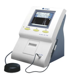
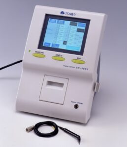
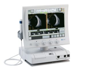
B Scan ultrasonography (USG) is a two dimensional  tool  with high frequency sound waves used for diagnosing lesions of posterior segment of the eyeball. It can accurately image intraocular structures and give valuable information of the status of lens, orbit, vitreous, retina, choroid, and sclera. However in many instances, it is used for diagnostic purposes even though pathology is clinically visible.

Perimetry

Perimetry is a standard method used in ophthalmology to assess patient’s visual field. It provides a measure of the patient’s visual function throughout their field of vision. A visual field defect is performed as soon as glaucoma is suspected. It evaluates vision loss due to glaucoma, damage to visual pathways of the brain and other optic nerve diseases.

Perimeter